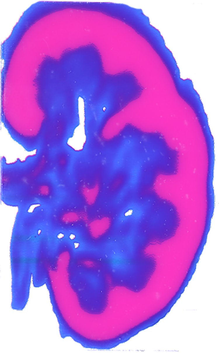
A group of College of Colorado researchers has developed a brand new technique for remodeling medical photographs, akin to CT or MRI scans, into extremely detailed 3D fashions on the pc. The advance marks an necessary step towards printing lifelike representations of human anatomy that medical professionals can squish, poke and prod in the actual world.

Voxel map of a cross part of a human kidney. (Credit score: Nicholas Jacobson)

Voxel map of a cross part of a human coronary heart. (Credit score: Nicholas Jacobson)
The researchers describe their ends in a paper printed in December within the journal 3D Printing and Additive Manufacturing.
The invention stems from a collaboration between scientists at CU Boulder and CU Anschutz Medical Campus designed to deal with a serious want within the medical world: Surgeons have lengthy used imaging instruments to plan out their procedures earlier than moving into the working room. However you may’t contact an MRI scan, stated Robert MacCurdy, assistant professor of mechanical engineering and senior writer of the brand new examine.
His group needs to repair that, giving medical doctors a brand new solution to print reasonable, and graspable, fashions of their sufferers’ numerous physique elements, right down to the element of their tiny blood vessels—in different phrases, a mannequin of your very personal kidney solely fabricated from delicate and pliable polymers.
“Our methodology addresses the important want to supply surgeons and sufferers with a larger understanding of patient-specific anatomy earlier than the surgical procedure ever takes place,” stated Robert MacCurdy, senior writer of the brand new paper and an assistant professor of mechanical engineering at CU Boulder.
The most recent examine will get the group nearer to attaining that aim. In it, MacCurdy and his colleagues lay out a way for utilizing scan knowledge to develop maps of organs made up of billions of volumetric pixels, or “voxels”—just like the pixels that make up a digital {photograph}, solely three-dimensional.
The researchers are at the moment experimenting with how they will use 3D printers to show these maps into bodily fashions which can be extra correct than these obtainable by means of present instruments.
The challenge, which is led by MacCurdy and CU Anschutz’ Nicholas Jacobson, is funded by AB Nexus, a grant program that seeks to spur new collaborations between the 2 Colorado campuses.
“In my lab we search for alternative routes of illustration that may feed, slightly than interrupt, the pondering strategy of surgeons,” stated Jacobson, a medical design researcher on the Inworks Innovation Initiative. “These representations turn out to be sources of concepts that assist us and our surgical collaborators see and react to extra of what’s within the obtainable knowledge.”
Slicing the orange
Human organs are difficult—made up of networks of tissue, blood vessels, nerves and extra, all with their very own texture and colours.
At the moment, medical professionals attempt to seize these constructions utilizing “boundary floor” mapping, which, basically, represents an object as a sequence of surfaces.
“Consider present strategies as representing a whole orange by solely contemplating the outside orange peel,” MacCurdy stated. “When seen that method, all the orange is peel.”
His group’s methodology, in distinction, is all juicy insides.
The strategy begins with a Digital Imaging and Communications in Medication (DICOM) file, the usual 3D knowledge that CT and MRI scans produce. Utilizing customized software program, MacCurdy and his colleagues convert that info into voxels, basically slicing an organ into tiny cubes with a quantity a lot smaller than a typical tear drop.
And, MacCurdy stated, the group can do all that with out dropping any details about the organs within the course of—one thing that’s inconceivable with present mapping strategies.
To check these instruments, the group took actual scan knowledge of a human coronary heart, kidney and mind, then created a map for every of these constructions. The ensuing maps had been detailed sufficient that they may, for instance, distinguish between the kidney’s fleshy inside, or medulla, and its outer layer or, cortex—each of which look pink to the human eye.
“Surgeons are continuously touching and interacting with tissues,” MacCurdy stated, “So we wish to give them fashions which can be each visible and tactile and as consultant of what they’ll face as they are often.”


